Hip Dysplasia was described in the dog in the year 1935. The difference between man and dog is that the dog's coxo-femoral Dysplasia is a hereditary disease, but it is not congenital: the dog is not born with Dysplasia, but due to the influence of environmental, dietary factors, excessive exercise, etc., coupled with an important genetic component, there is an imbalance between muscle mass and skeletal development, resulting in a lack of congruence between the acetabulum and the head of the femur . Basically we have two types of Dysplasia.
1 - Acetabular – Typical in the German Shepherd and Labrador Retriever, where there is flattening of the acetabulum associated with a low acetabular coverage of the femoral head.
2 - From the neck of the femur – It is characterized essentially by the alteration of the femoral angle and the lack of pressure at the level of the acetabulum. Loss of contact between the femoral head and the acetabulum causes joint instability and coxo-femoral laxity, subsequently leading to osteoarthrosis. Classically affects large and giant breeds. It has been described in more than 70 breeds. The incidence is 48% in S. Bernardos, 31% in Bullmastiff, 23% Golden Retriever, 22% Rottweiller, 21% German Shepherd. It is described in small breeds such as the Cocker Spaniel and even in cats (Persian). It affects bilaterally in 90% of the cases, and there is no sexual predisposition.
Genetic Factors – The heredity of this disease has a polygenic character (it is not known how many genes intervene). Many animals may show a normal phenotype with correct radiography, but they are genotypically carriers of the dysplastic character and transmit to offspring, which greatly complicates their eradication. The constitution of the breed itself is also a determining factor for the presentation of Dysplasia, asynchrony between bone and muscular development (Ex: Labrador), intrinsic morphology of the shallow acetabulum in the German Shepherd, more concave in the Boxer, the angulation and orientation of the head femur in the Mastin and Pyrenean Mountain, and the typical joint laxity of the German Shepherd, explain the different percentages of incidence in similar weight and weight development breeds.
Environmental factors – An excess of feeding. Bound, usually, to feed "ad libitum" influence the speed of growth of the animal. A young animal with bone architecture that is not yet compact, which has to bear an excessive weight may be willing to develop Ankle Dysplasia. Overeating with diets high in energy calcium, vitamins, etc., should be avoided especially at the maximum age of growth, between 3 and 8 months of age.
Exercise – Factor to Consider in the Etiopatology of Hip Dysplasia. Violent exercises increase joint laxity. A moderate exercise that allows proper muscle development increases joint stability and helps prevent hip dysplasia. Clinical symptoms are many varying and with a certain independence of radiologically evident osteoarthritic lesions. There is no parallelism between clinical symptoms and radiographic signs, presenting a wide range of possibilities of these asymptomatic dysplastic animals to paraplegic animals. An abnormal gait, joining the curvilines, difficulty in getting up or jumping, pain in the manipulation of the extremity especially in hyper extension, etc., are signs suggestive of Dysplasia that will have to be confirmed with a radiology.
Young animals of 6 to 12
months of age that manifest signs of more or less
intermittent and transient forms. This is due to the
existence of micro fractures. Painful, which occur
mainly on the acetabular edge due to joint laxity.
Subsequent rapid ossification leads to a more or less
transient disappearance of pain.
A second group of animals would be represented by
animals 4 to 5 years or older when irreversible
osteoarthrosis lesions have already been established. In
young animals, we can try to perform an early diagnosis
of joint laxity using the Ortolani test. The animal is
placed in lateral decubitus, the knee is pressed towards
the trochanter which facilitates the luxuation of the
femoral head. Keeping the pressure, we move the
extremity towards the outside (abduction), which will
cause the head of the femur to be repositioned inside
the acetabulum. In dogs with joint laxity
(predisposition to Dysplasia), we notice a noise when
the femoral head recovers its normal position.
The value of the diagnosis is debatable, since if it
is positive the animal will usually have Dysplasia, if
it is negative the doubt persists.
The Bardens sign is also used to determine early excess
articular laxity in puppies. It consists of trying to
separate the femoral head from the acetabulum by a high
abduction force, with the animal positioned in lateral
decubitus. About 75% of the animals positive for these
tests will be dysplastic in adulthood.
Radiological study is currently the only means of diagnosing coxofemoral Dysplasia. In the German Shepherd, for example the reliability of radiographic detection is 16% at 6 months of age, 70% at one year of age, 82% at 18 months of age and 95% at 2 years of age. The official radiograph should be done at 18 months (never in females with heat). The standard radiological technique, accepted universally, requires the sedation or anesthesia of the animal, placing the animal in the supine position with the posterior distended, parallel and subjected to internal rotation so that the kneecaps are located on the trochlea of the femur, avoiding the rotation of the animal. pelvis. The symmetry must be perfect.
The classification of Dysplasia degrees varies by country. The classification accepted in our country is the one proposed by the International Cynological Federation (FCI):
|
No Signs of Dysplasia |
Grade A |
|
Transition form |
Grade B |
|
Mild Dysplasia |
Grade C |
|
Moderate Dysplasia |
Grade D |
|
Serious Dysplasia |
Grade E |
In order to make the correct reading of the radiography, we use lame metric measurements:
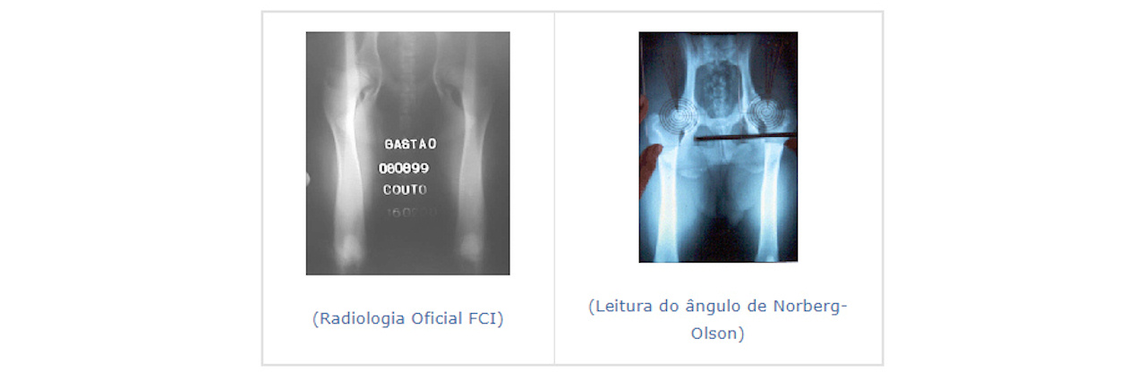
Norberg-Olson Angle
Cervical-Diaphyseal Angle
Retro and anteversion angle
Acetabular Coverage
Conservative Treatment – Prevents the prevention
of joint cartilage injury in the young dog and the
relief of pain secondary to arthrosis in the adult dog.
Rest, weight reduction and controlled use of anti-inflammatories.
Surgical – The surgical
techniques of Ankle Dysplasia are intended to suppress
pain or correct bad joint formation
Pectinectomy – The pectin
muscle acts as an adulterer. The contraction of this
muscle in a dysplastic hip predisposes to subluxation
and increases pain. A tenotomy or tenectomy of pectinium
can achieve a short-term anti-pain effect, but does not
modify the progression of osteoarthrosis signs. Most
patients return to clinical signs in the near future
depending on the age and degree of osteoarthritis
present at the time of surgery. In very specific cases
we recommend this surgery.
Triple osteotomy – The
triple osteotomy of the pubis, ischium and ilium,
releases the acetabular part, so that it can be
reoriented, achieving a greater congruence of the same
with the femoral head, thus decreasing the laxity of the
joint capsule, and avoiding lesions degenerative
diseases of the joint. The ideal candidate for a triple
osteotomy is an animal with acetabular Dysplasia of 7 to
12 months of age with articular laxity that clinically
manifests symptomatology and that does not show signs of
joint degeneration. We can rotate the acetabulum 20, 30
or 40 degrees.
The Veterinary Hospital of Oporto with a surgical
unit headed by Dr. Mário Santos, has been performing the
triple osteotomies in many affected animals for 4 years,
due to this disease in all regions of the country. We
believe that it is the best surgical method to correct
Dysplasia, being the selection of patients, as well as
age, of primary importance for success to be 100%.
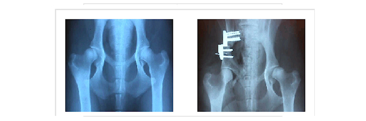
The rotation in the
case described above was 30 °, the radiography was
performed after surgery.
We can verify the total introduction of the femoral head
into the acetabulum after the surgical procedure.
Triple osteotomy – It is the most recent surgery
that we use to correct Dysplasia in animals that have
already passed the 12 months of age and that can no
longer perform a triple osteotomy. However, they are not
candidates for hip prosthesis surgery, much less an
excision arthroplasty.
The surgery consists of raising the acetabulum with a
very effective technique that keeps the head of the
femur stabilized.
We have been performing this technique for two years,
achieving extraordinary results..

Excision of the femoral head should only be used as a last resort. The end we pursue is the suppression of pain, eliminating the joint. Functional recovery is slower.

The replacement of the
femoral hip joint in animals with osteoarthrosis by a
prosthesis is an increasingly used technique.
Usually cemented prostheses with a success rate of 60 to
100% are used depending on the technique used.

Source: Boxer Club
of Portugal website.
PORTUGUESE ASSOCIATION OF VETERINARY PHYSICIANS
» Ankle Dysplasia Control Program
PORTUGUESE ASSOCIATION OF VETERINARY PHYSICIANS